7 QUESTIONS ABOUT SPINAL SUBARACHNOID “CYSTS”
Jay McDonnell, DVM, MS, Diplomate ACVIM (Neurology)

1. What is a spinal arachnoid cyst?
A spinal subarachnoid cyst is not actually a cyst; instead it is a fluid-filled dilation within the subarachnoid space. It’s best referred to as an Arachnoid Diverticulum (plural=Diverticula). The term “pseudocyst” has been used to describe this abnormality as well.
2. What are the clinical signs and history of a Spinal Arachnoid Diverticulum?
It depends on the location. If found in the thoracolumbar spine, spastic long-strided pelvic limb weakness with spinal ataxia that is slowly progressive over several months is one clinical sign. Fecal incontinence is another common sign with thoracolumbar arachnoid diverticulae. Anal tone is normal; however dogs will drop stool in the house or outside while walking (without posturing and without apparent awareness).
3. Who is affected by an Arachnoid Diverticulum?
Any dog can be affected; however middle-aged Pugs and juvenile to young adult Rottweilers are over-represented. Arachnoid diverticula have also been reported in cats, but is relatively rare.
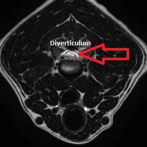
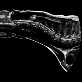
4. How is an Arachnoid Diverticulum diagnosed?
MRI (like the ones shown below) is the safest and most complete way to evaluate for this spinal anomaly; however, CT coupled with myelography can be used, as well.


5. What causes an Arachnoid Diverticulum?
The cause is unclear; however, it is suspected to be congenital in most young animals, such as Rottweilers. Abnormal tissue within the arachnoid trabeculae likely interrupts normal pulsatile flow of CSF. This abnormal tissue may be meningeal fibrosis or congenital in nature. In Pugs and other brachycephalic breeds, it has been suggested to be secondary to a chronic vertebral instability related to hypoplastic facets. We are unsure if the instability is the cause or if the boney anomaly is associated with a concurrent congenital anomaly of the spinal cord itself (diverticulum).
6. What is the treatment?
Surgery is the best treatment and involves a laminectomy, dural marsupialization, breakdown of adhesions (if present), +/- surgical stabilization if indicated.
7. What is the prognosis with and without treatment?
The prognosis is unknown; however the majority of dogs improve initially after surgery. There is a risk of recurrence of the cyst and some dogs have been known to undergo a secondary surgical procedure.