
VETERINARY MRI & CT ADVANCED IMAGING IN HUNT VALLEY, MD
CT and MRI are essential medical imaging techniques that offer detailed insights into a patient’s body. VNIoC provides advanced diagnostic imaging for pets and referring veterinarians, supported by exceptional image quality and dedicated veterinary anesthesia care.
Use this page to learn what to expect, compare CT vs. MRI scans, and request outpatient imaging for your patient or pet.
Advanced Veterinary Imaging for Pets
Advanced veterinary imaging uses technologies such as MRI and CT to create detailed images of a pet’s brain, spinal cord, joints, bones, organs, soft tissues, and internal structures. MRI is often preferred for brain, spinal cord, joint, and soft tissue conditions, while CT is commonly used for bones, trauma, certain tumors, internal organs, and vascular structures.
WHAT TO EXPECT DURING YOUR PET’S MRI OR CT SCAN
-
![]()
01
Check-In and Consent
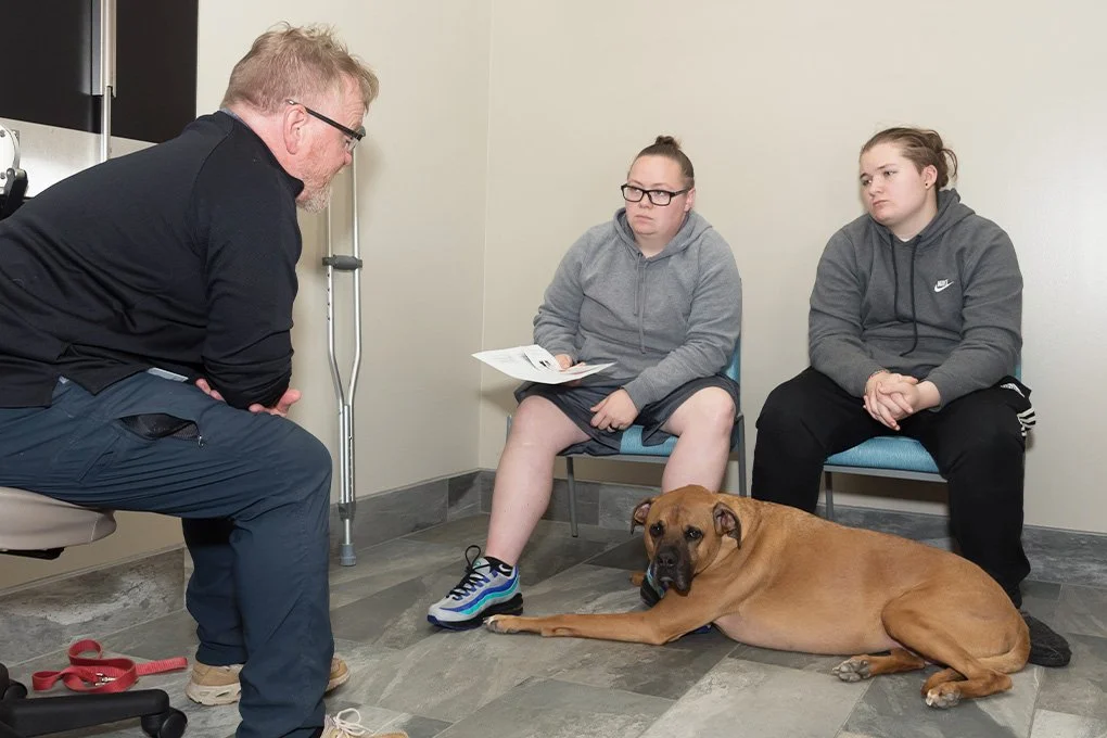
Your pet is checked in, consent forms and fasting procedures and medications are reviewed, and our team confirms the imaging plan before the procedure begins.
Your pet will be with us all day — expect them to be discharged in late afternoon.
-
![]()
02
Scan Preparation
Our staff prepares your pet for the scan. They follow anesthesia or sedation protocols based on the patient’s needs to make sure they don’t experience stress or discomfort.
-
![]()
03
MRI or CT Imaging
Your pet receives the appropriate scan. CT scans are usually faster, while MRI scans often take longer due to soft tissue detail.
-
![]()
04
Recovery and Monitoring
After imaging, your pet is monitored during recovery by the veterinary team.
-
![]()
05
Results and Next Steps
Imaging findings help guide the diagnosis, treatment plan, and next recommendation
Most pets are discharged late afternoon. A neurologist will speak with you about your pet at pick-up time if they are available. They may also want to consult a radiologist. Parents will receive a clinical summary email after their pet’s visit.


CT vs. MRI for Pets: What’s the Difference
The choice between CT and MRI depends on the clinical scenario, the body part being examined, and the information required by the veterinarian to make an accurate diagnosis.
CT Scan
Best for: Bones, trauma, some tumors, organs, and vasculature
Scan time: Typically faster, often 10-20 minutes
Radiation: Uses X-rays
Anesthesia: May require sedation or anesthesia
Image type: Cross-sectional and 3D images
MRI Scan
Best for: Brain, spinal cord, soft tissue, and joints
Scan time: Typically longer, often 40-60 minutes
Radiation: No ionizing radiation
Anesthesia: Usually requires anesthesia
Image type: High-resolution soft tissue images
Veterinary CT Scans for Bones, Trauma, Tumors, Organs, and Vasculature
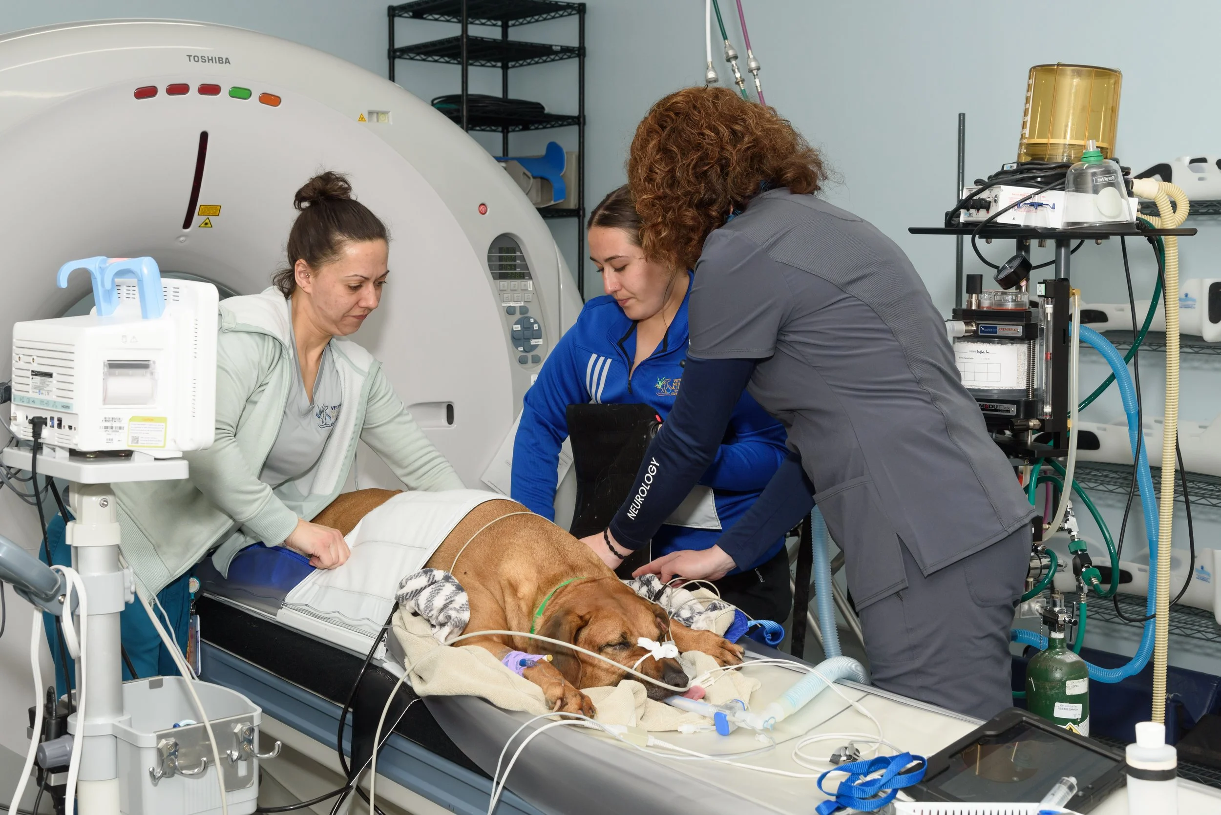
CT scanners use X-rays to provide cross-sectional and 3-dimensional images of the area of interest, making CT useful for evaluating bones, certain tumors, internal organ structures, and vasculature.
What a Veterinary CT Scan Shows
CT can provide detailed views of bone structures, internal organs, tumors, and vascular structures.
When CT May Be Recommended
Bone injuries or fractures
Trauma assessment
Certain tumors
Internal organ and vascular evaluation
Veterinary MRI for Brain, Spine, Joint, and Soft Tissue Conditions
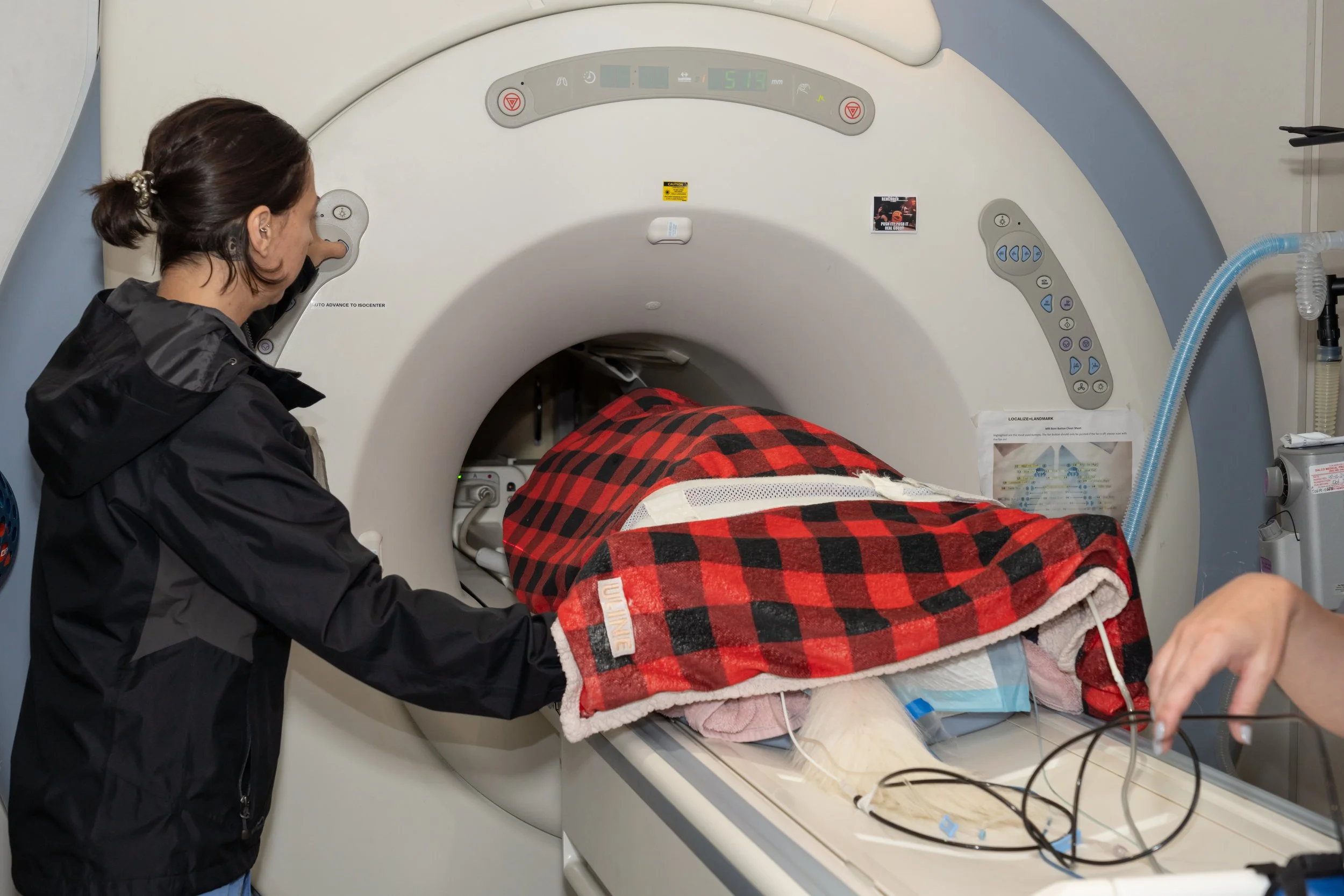
MRI uses a powerful magnetic field to produce high-resolution images of soft tissues. It is often considered the gold standard for imaging the spinal cord and brain, and it can also provide detailed joint images.
What a Veterinary MRI Shows
MRI can show detailed soft tissue structures, including the brain, spinal cord, joints, and certain musculoskeletal conditions.
When MRI May Be Recommended
Brain and spinal cord evaluation
Soft tissue detail
Joint and musculoskeletal disorders
Neurological conditions that may not be visible on radiographs
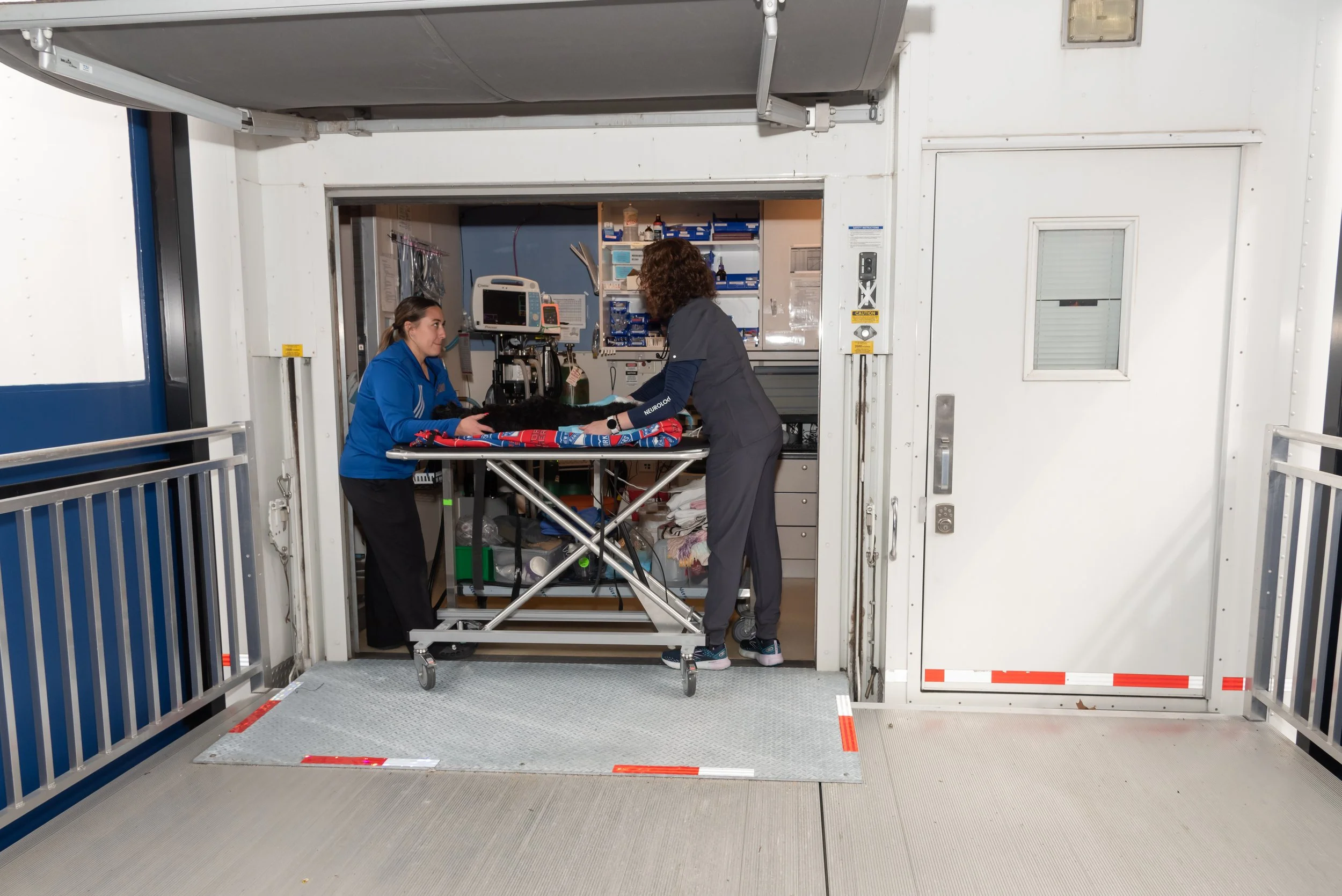
Choosing the Right Veterinary Imaging for Your Patient
Generally, MRI is superior for visualizing soft tissue, while CT provides excellent detail for bone structures. The right choice depends on the suspected condition, the patient’s stability, and the diagnostic information needed.
When MRI Is Usually Preferred
MRI is often preferred for brain lesions, spinal cord conditions, non-calcified disc extrusions, soft tissue tumors, and intraparenchymal lesions.
When CT Is Usually Preferred
CT may be preferred for acute head trauma, skull fractures, acute hemorrhages, bony lesions, fractures, discospondylitis, bony neoplasia, and calcified disc extrusions.

Veterinary Anesthesia Safety During MRI and CT Scans
We prioritize the comfort and safety of your pet during their procedure. Our imaging department veterinarian, Dr. Elana Rybak has extensive experience in anesthesia, ensuring the highest standard for our patients. Throughout your pet’s procedure, one of our experienced veterinarians will be specifically dedicated to managing your pet's anesthesia— ensuring their comfort and safety every step of the way:
Pre-operative Assessment
Each patient’s health status is evaluated before imaging to determine the most appropriate anesthesia approach.
Anesthesia Administration and Monitoring
During MRI and CT scans, anesthesia is carefully administered and monitored by experienced veterinary professionals.
Post-Operative Recovery
Patients are supervised during recovery and monitored for comfort and safety.
Refer a Patient for Outpatient Veterinary MRI or CT Imaging
With advanced MRI and CT scanners, VNIoC provides image quality that supports accurate diagnosis and treatment planning. The goal is to support timely imaging services and improved patient outcomes.
Veterinary MRI and CT Scan FAQs
-
CT uses X-rays to create cross-sectional and 3D images.
MRI uses a powerful magnetic field to produce detailed images of soft tissues.
-
CT and MRI images show different types of detail.
CT is useful for bones and certain internal structures, while MRI is useful for soft tissue, brain, spinal cord, and joint detail.
-
CT scans are typically faster, often 10-20 minutes. MRI scans typically take longer, often 40-60 minutes.
-
Anesthesia or sedation needs depend on the scan type, patient health, and the need for the pet to stay still during imaging.
-
Cost depends on the type of scan, body area, anesthesia needs, contrast use, and case complexity. Contact the team for guidance based on your pet or patient’s needs.
-
MRI and CT can help evaluate neurological, orthopedic, soft tissue, internal organ, tumor, trauma, vascular, and spinal conditions depending on the scan type.
-
Metallic implants may affect MRI safety or image quality. The team should review the implant type and clinical situation before imaging.and safety concerns.
-
Contrast may be recommended depending on the suspected condition and the diagnostic information needed.
-
CT uses X-rays, so it involves radiation exposure. MRI does not use ionizing radiation.
-
Potential risks depend on the scan type, anesthesia or sedation, contrast use, and the patient’s health. The team reviews these considerations before the procedure.
Trusted Advanced Veterinary Imaging
in Hunt Valley, MD
Local Specialty Care
Advanced veterinary imaging services for pets and referring veterinarians in Hunt Valley, Baltimore, and surrounding Maryland areas.
Dedicated Anesthesia Monitoring
Imaging procedures are supported with veterinary anesthesia monitoring and recovery oversight.
Referral Support
Clear CT and MRI request paths help referring veterinarians submit outpatient imaging requests efficiently.
Request Advanced Veterinary Imaging
Request outpatient CT or MRI imaging, or contact the team with questions about advanced imaging for your pet or patient.