
An 11 year old MN Lhasa Apso was referred with a three month history of recurring swelling and discomfort over the left submandibular region. Over this time period, three separate exploratory surgical procedures had been performed on this area at different hospitals. The last procedure had been performed within a week of referral, with a rapid return of the swelling and discomfort postoperatively.
In that procedure, the surgeon had removed the mandibular salivary gland as well. Histopathology and cytology reports from each of the procedures described non-specific pyogranulomatous inflammation, cellulitis, with some atypical histiocytic cells. This inflammation was also associated with the left mandibular salivary gland. No tissue culture was submitted from samples obtained during the last surgery. Vimago HD 3D CT revealed soft tissue swelling and an irregular boney proliferation at the left mandibular angular process, along with bilateral otitis media and externa. A small sequestrum was noted adjacent to the angular process.
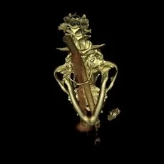
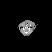
Fine needle aspiration of the soft tissue swelling was submitted for aerobic culture, with no bacterial growth obtained. Recommendations included surgery to remove/culture the sequestrum and biopsy the angular process. This fourth surgical procedure was declined by the owner, who subsequently requested non-surgical treatment. Oral enrofloxacin and amoxicillin/clavulinic acid were begun. Phenobarbital was also begun in an attempt to inhibit saliva production. Topical antibiotic/anti-yeast treatment for otitis externa was provided bilaterally.
The patient was treated with adjunctive one-hour BID HBOT sessions at 1.5 ATA over a 7 day period, along with daily Artemis NRT Laser therapy over the lesion site at (150mj/30Hz) for 3 minutes and (250mj/15Hz) for 5 minutes.
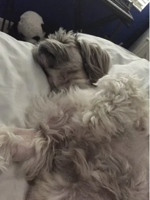
Within 24 hours after commencement of this regimen the mandibular swelling was markedly reduced, and completely resolved over the next week. The patient was discharged on phenobarbital, enrofloxacin, and amoxicillin/clavulinic acid. The phenobarbital was weaned after the first two weeks of therapy, and the antibiotics were continued for a total of 6 weeks.
The owner declined follow-up Vimago CT at 8 and 10 weeks after the initial Vimago CT, in order to re-evaluate the lesions initially recognized on the angular process of the mandible. The owner was pleased with the results, and stated that her pet was clinically normal one month after discontinuing all medications.
The owner was willing to provide digital images of the mandibular area, which were obtained at 10 weeks after initial presentation (4 weeks after the cessation of the medications). The mandibular area appeared normal, with the exception of a small cutaneous scar remaining where the surgical drains had been placed. (Image above left)
Adjunctive HBOT has been found to be beneficial and effective in wound healing in evidenced-based studies. (Ref: Goldman RJ, “Hyperbaric Oxygen Therapy For Wound Healing and Limb Salvage: A Systemic Review”, Physical Medicine and Rehabilitation. 2009:1:471-489).
© RKLyman, LLC. All Rights Reserved.