
A Determined Dachshund Defies Meningoencephalitis of Unknown Etiology (MUE)
When five-year-old Louie, an adorable Dachshund, started having episodes of abnormal movement in September 2024, his parents were understandably alarmed. Louie would first lift his back leg, then his front, before falling over and shaking. He also showed signs of severe neck pain — which is never a good sign in a long-backed “sausage dog.”
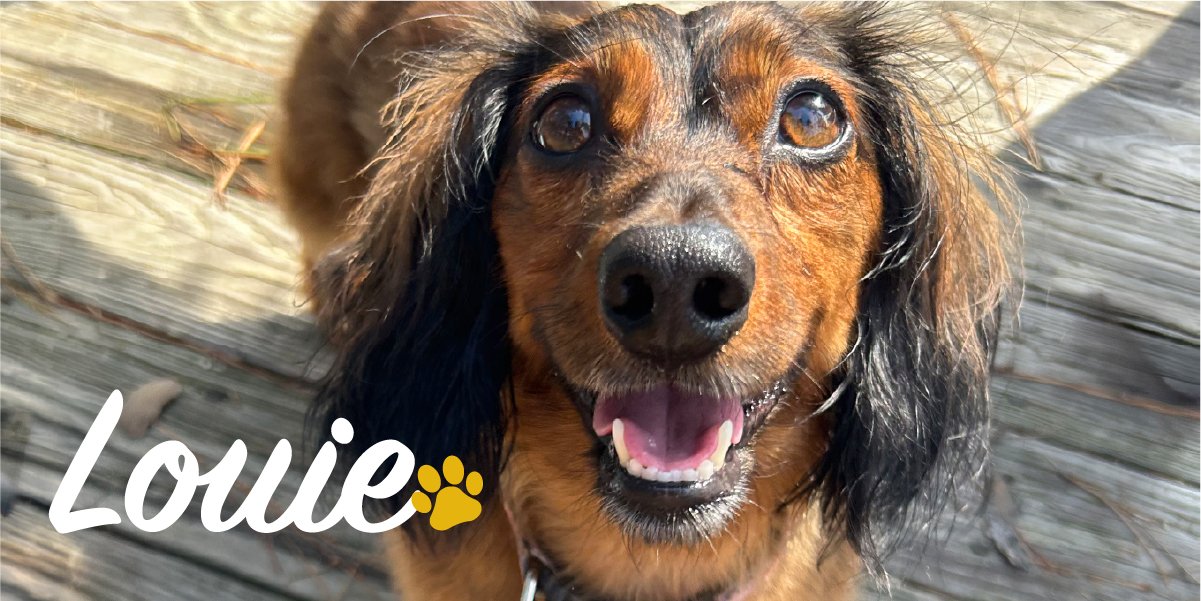
“His condition started as a series of frightening, unexplained episodes that came completely out of nowhere. We initially thought Louie was having seizures. He would suddenly fall over, lose his balance, start shaking, and become completely disoriented,” Louie’s parents told us. “Watching him go through these episodes was absolutely terrifying, and we had no idea what was happening to our sweet boy.”
Louie’s parents brought him to Veterinary Neurology & Imaging of the Chesapeake (VNIoC) where Dr. Julianna Mannix examined him. She confirmed the neck pain and noted delayed proprioception in all four legs, meaning that Louie didn’t know where his paws were in space. Concerned that these episodes might be paroxysmal dyskinesia rather than seizures, Dr. Mannix recommended an MRI to help make a definitive diagnosis.
Diagnosing MUE with MRI Imaging & Labs
Louie’s MRI showed inflammation in multiple locations throughout his brain and spinal cord, but his infectious disease testing was negative, ruling out viral, bacterial, fungal, or protozoal encephalitis. Because Louie’s intracranial pressure was high, a spinal tap for cerebrospinal fluid (CSF) analysis could not be safely performed. Dr. Mannix diagnosed him with Meningoencephalitis of Unknown Etiology (MUE). Clinical Note: While a definitive confirmation of MUE requires an invasive brain tissue biopsy, the associated risks and financial implications often lead to a presumptive diagnosis based on imaging and labs.
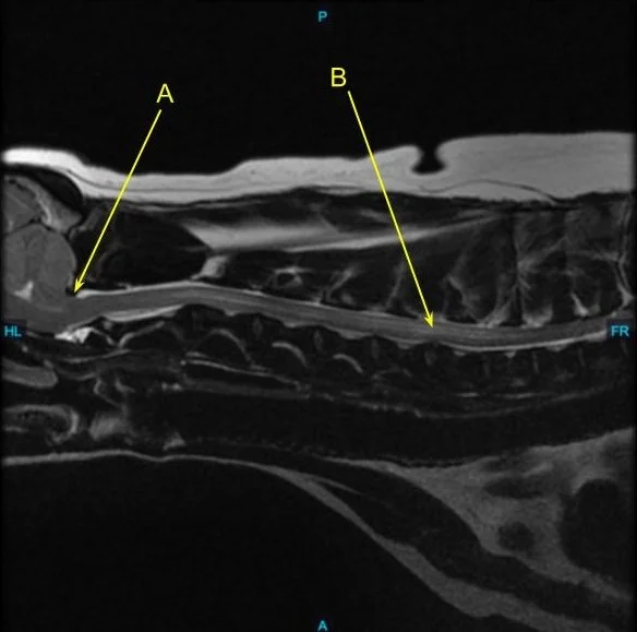
This side view of Louie’s cervical spinal cord shows: A.
On the back of the skull, p
art of his brain is partially herniating through the opening from the skull into the spinal canal indicating increased intracranial pressure. B. The bright line within the spinal canal is most consistent with inflammation extending from the brain.
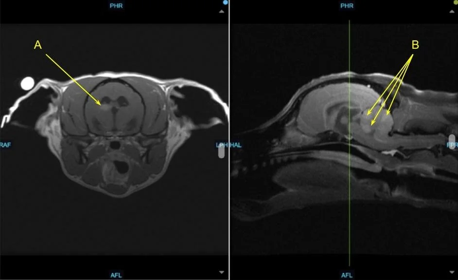
These images of the brain show: A. The right, contrast enhancing lesion within the brain consistent with inflammatory disease. B. Multifocal, bright, contrast enhancing lesions within the cerebellum consistent with inflammatory disease.
In dogs affected by MUE, the immune system mistakenly attacks healthy tissue within the brain and spinal cord. Immune cells enter the central nervous system and release inflammatory chemicals that are intended to fight disease, but instead damage normal nervous tissue. Symptoms vary depending on where the brain and/ or spinal cord are affected, but can include seizures and muscle tremors consistent with Louie’s episodes of instability.
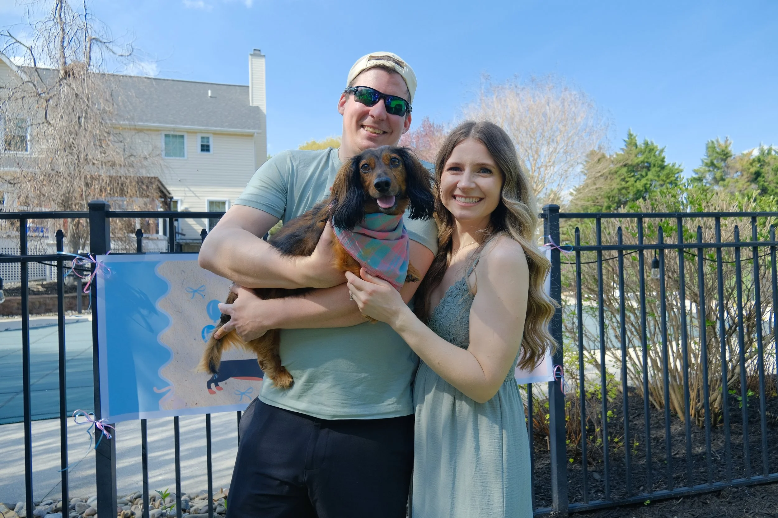
Louie the Dachshund with his parents
Treating MUE in Dogs with Cytosar
Louie was treated with prednisone and Cytosar, an injectable chemotherapy medication prescribed for severe brain inflammation in dogs, Dr. Mannix also prescribed Keppra to help manage hisepisodes. Little Louie proved to be a champ, responding so well to treatment that Dr. Mannix stopped his Cytosar treatments and he was able to continue on prednisone alone.
MUE Prognosis and Long-Term Management
As of his July 2026 check-up appointment, just shy of the two year anniversary of his first visit to VNIoC, Louie was doing extremely well. He is neurologically normal on a very low dose of prednisone, with a plan to taper off Keppra.
Although clinical outcomes vary based on the specific pathology, patients demonstrating a positive therapeutic response beyond the six-month mark often carry a more favorable long-term outlook compared to those who fail to stabilize during the initial phase of intervention.
Louie’s success is especially meaningful because MUE was once associated with a very poor prognosis, with many cases becoming terminal within just a few months. Thanks to modern, intensive therapies, success stories like Louie’s are becoming far more common—giving beloved pets more quality years with their families.
“Dr. Mannix never gave up on Louie. Because of the care, expertise, and dedication he received, Louie is doing absolutely wonderful today. He is back to being his playful, loving, happy self, and we are so incredibly grateful that we get to continue making memories with him!
Louie isn’t just our dog—he is a huge part of our family. And now, he is about to become a big brother to his first human sibling, which is something we never could have imagined during those scary days when we weren’t sure what his future would hold.
We truly cannot thank Dr. Mannix enough for everything she did for Louie and for our family!” — Louie’s parents