
Paralyzed to Full Recovery: A Pawrent’s Perspective
“We won the lottery with her,” says Jessica of her family's 10 year old Jack Russell Terrier mix, Harley. “She loves her people, and to be in the woods with me going for a hike. She's very playful and energetic and then when it's time to snuggle, she's such a snuggle bug.”
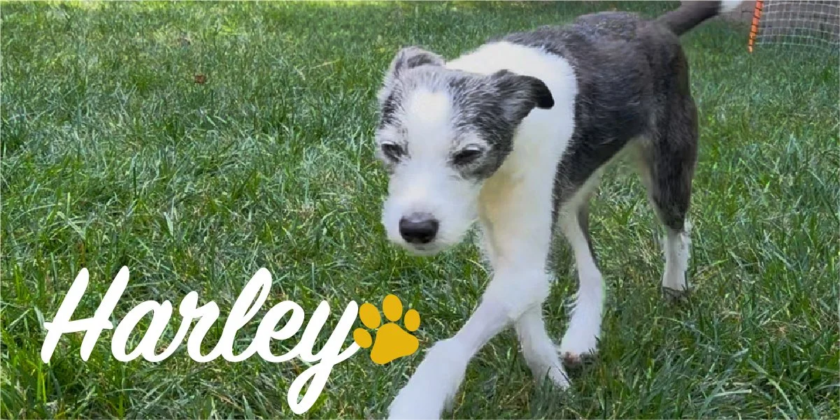
Then over a May weekend, Harley went from active to paralyzed.
Jessica continues, “One Friday afternoon, Harley ran up to the fence and barked at the lawnmower on the other side–and then yelped. We had no idea what had happened to her. As the evening progressed, it was clear that she was in a lot of pain. We thought for sure it was in her back. She started to get very stiff and by Saturday morning, she was basically paralyzed. It was crazy how fast it happened. So we brought her to Pet+ER.”
“The vet there was fantastic. They gave us a steroid and a pain pill and said she should be better in 24 to 48 hours. We wanted to give her the full 48 hours and see, but it was very clear by Monday morning, she wasn't getting any better. It was absolutely terrifying—especially for our kids—to watch the family dog that we've had for nearly a decade struggle like that.”
After checking back in at Pet+ER, Harley was immediately referred to VNIoC for a neurology consult.
“From there it's kind of a blur. The tech, who was lovely, asked us what happened and she had to wait for probably 10 minutes for me to be done crying to answer. When Dr. McDonnell came in, I showed him some of the videos of how Harley had been acting and her lack of ability to walk. He was so kind with her and showed such compassion that we knew we were in the right place. After he examined her, he said he thought it was pretty clear that it was something in her neck. He advised us to get the MRI and we would go from there, but very likely, she would need surgery.”
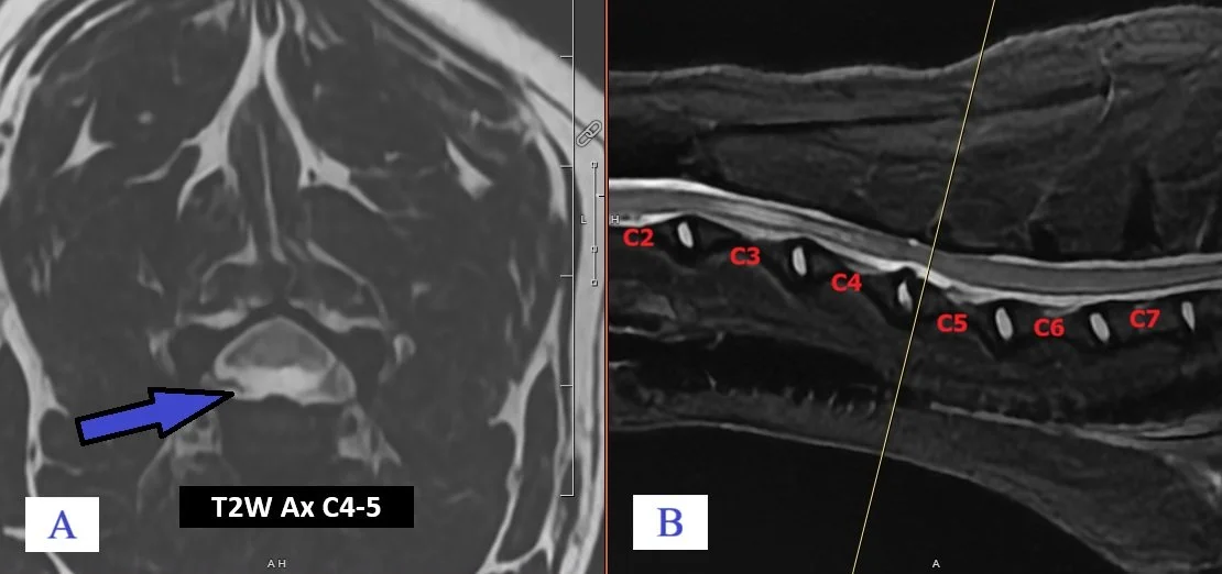
Figure 1A: Axial T2W image at the level of C4-5 which shows a large hydrated disk that ruptured dorsally into the spinal canal and severely compresses the spinal cord. This is a hydrated nucleus pulposis extrusion (HNPE).
Figure 1B: Mid-sagittal STIR image of the spinal cord with the cervical vertebrae lableled in red. Harley’s head is to the left. The line between C4-5 is at the level of the axial image (A).
[On the MRI, large volume of lobulated extradural material spanning the C4-C5 intervertebral disc space resulting in ventral compression to the cord. These imaging findings were thought to be a hydrated nucleus pulpous extrusion (HNPE) causing concurrent compression of the cord.]*
“Dr. McDonnell was so confident that, with [ventral slot] surgery, she would have a good prognosis. He said, ‘I honestly feel like she'll be hiking with you again in the fall.’
“The surgery went extremely well. VNIoC took amazing care of her. After we got her home, we were nervous but everything was explained to us very well and everybody at VNIoC was just so supportive.
“Six weeks after the surgery, you would not even notice that she has anything wrong. Every once in a while, she has maybe a little challenge with her balance, but that's very rare. The hard part now is keeping her settled down!”
“I don't think she would be with us without Dr. McDonnell and what he did for her. It was just incredible.”
Contributing to Harley’s positive outcome were her parents who watched her closely and did everything right. Thanks to their prompt actions and continuing follow-up, Harley was a great candidate for corrective surgery and had a good prognosis.
*An HNPE differs from other disc herniations because the material extruded into the spinal canal is gelatinous or liquid, rather than solid or calcified.
The clinical presentation of HNPE also varies from classic acute intervertebral disc extrusion (IVDE) in several key ways. Classic IVDE typically affects young to middle-aged, small, chondrodystrophic or toy breeds, whereas HNPE is more common in middle-aged to older medium and large breed dogs. While both conditions cause discomfort, dogs with IVDE often exhibit more intense pain and cervical rigidity. Conversely, dogs with HNPE usually present with more severe neurological deficits.
Although clinical signs serve as helpful indicators for estimating the likelihood of a specific disk type, they should not be used to establish a definitive diagnosis. MRI or surgical exploration is required to definitively distinguish between HNPE and IVDE.