Emergency IVDD Surgery in Dogs: Misty’s Three-Site Hemilaminectomy Success Story
Misty was rushed to Pet+ER on the Saturday before Valentine’s Day when she developed excruciating back pain and weakness in her rear legs. At the initial presentation, Misty was able to walk without help but by Sunday, she was nearly paralyzed, so Pet+ER brought in Dr. Jay McDonnell and the on-call VNIoC team to help Misty before her condition worsened.
Dr. McDonnell determined that Misty was experiencing symptoms consistent with intervertebral disc disease (IVDD), a condition commonly referred to as a “slipped” or ruptured disc in dogs. Misty then underwent an emergency spinal MRI — the gold standard for diagnosing IVDD and determining whether surgical decompression is required.
Misty’s MRI of her thoracolumbar spine revealed an extruded disc at L4-5 with a large amount of disk material spanning across her spinal cord from vertebrae L3-4 to L5-6. This material and the compression it caused on the spinal cord were responsible for the increasing weakness in Misty’s hindlimbs. It was clear that surgery was necessary and urgent.
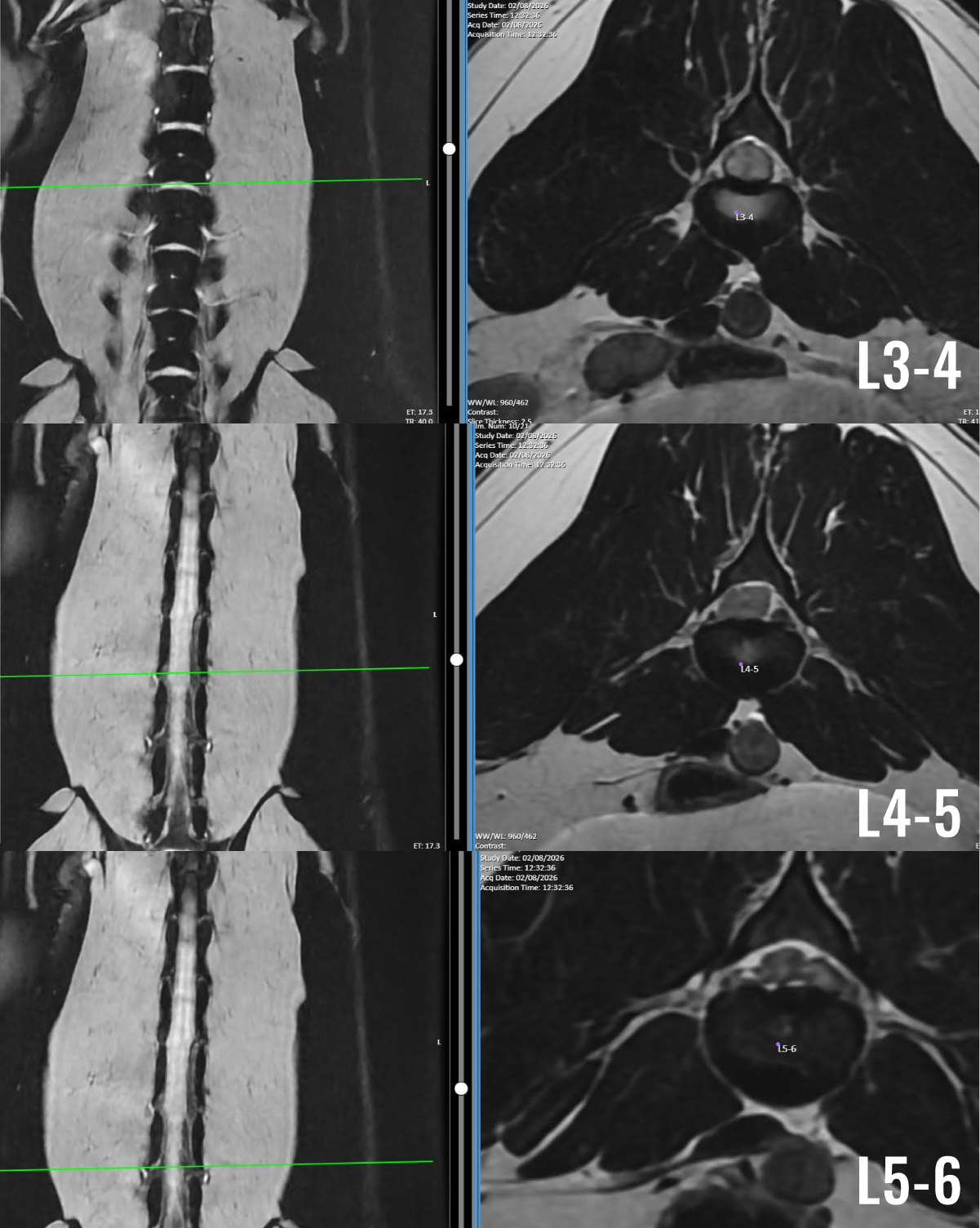
MRI images show Misty’s spinal cord compression and disc material at each site (L3-4, L4-5, L5-6). These MRI images demonstrate severe spinal cord compression caused by extruded intervertebral disc material.
Given the severity of her spinal cord compression, Dr. McDonnell performed a three-site hemilaminectomy (a surgical procedure that removes bone to relieve pressure on the spinal cord). After surgery, Misty showed immediate improvement. She was hospitalized for two days, and made steady improvements. Post-operative care following hemilaminectomy for IVDD is critical to recovery and typically includes:
24-hour monitoring and care, including medication administration, feeding/watering, monitoring of IV medications/fluids, and slow leash and sling walks outside.
Laser therapy over the muscles on either side of the spinal cord around the surgical sites.(In Misty’s case, this was the muscles on either side of her L3-4, L4-5 and L5-6 vertebrae).
Passive Range of Motion (PROM) exercises and other rehabilitation exercises, like a peanut ball and stretching.
Over her two days in hospital, Misty recovered more and more. She returned home with her pawrents, who received detailed post-op care and rehab instructions from the VNIoC team. A little over two weeks after discharge, Misty was back to her normal, tail-wagging self.
Like many dogs undergoing timely surgical treatment for IVDD, Misty’s outcome was excellent. Early diagnosis, advanced imaging, and prompt hemilaminectomy surgery gave her the best chance at a full recovery.
Check out Misty’s progress: